Our group employs a multidisciplinary approach including analytical methods, physics models, algorithms, computer simulations and instrumentation to problems in the area of imaging physics. Applications of this research can be in the areas of medical imaging (such as early cancer deetction), biology, material sciences, defence&security and biophysics. Some of the ongoing projects are listed below. Further information for prospective postdoctoral fellows, graduate students, undergraduate students or high school interns can be found here.
Research key words: Imaging Physics, Spectral x-ray imaging, Bio-photonics, Medical imaging, Tomography, Photon counting detectors, Tomographic reconstruction thods for sparse data, Image science, Image perception, Optics, multi-modality imaging and Biophysics.
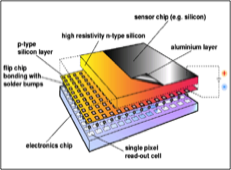
Spectral X-Ray Detection: Current X-ray Imaging in clinical setting utilizes an energy integrating detector along with an incoherent polychromatic x-ray tube. There has been a recent surge in research and technology development related to the use of photon counting spectral x-ray detectors for x-ray imaging. The ability to separate detected x-rays based on their energy would open new avenues of quantifying multiple complimentary object properties and hence enable contrast enhancement using x-ray imaging.
Our group is exploring these options with industrial and academic collaborators such as via Medipix collaboration (CERN, Geneva) to utilize Medipix2 and Medipx3 detectors for medical imaging applications. Besides exploring innovative applications, we are also involved in investigating correction and calibration strategies for future utility of these spectral detectors. This involves laboratory experiments, experiments at synchrotron facilities, modeling of detector physics and development of algorithms.
Phase Contrast X-Ray Imaging: Current Clinical X-Ray imaging relies of attenuation contrast between various tissue types which leads to poor soft tissue contrast. In breast imaging, attenuation of dense breast tissue and cancerous tissue is negligible leading to poor visibility of cancers in dense breast particularly during early stage of their growth. Phase contrast x-ray imaging is being investigated as an alternate contrast mechanism.
High radiation dose and imaging time resulting from cumbersome and multiple measurements required in the current phase contrast imaging methods have been a major hurdle in their clinical translation. Our group is exploring novel and single-step spectral phase contrast imaging methods with the goal of reducing radiation dose while estimating x-ray absorption and phase imagery with quantitative accuracy.
This work involves theoretical developments, computer simulations and laboratory prototype development.
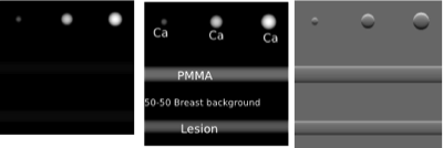
(Left to right): Absorption, phase and differential phase of signals in a breast background. Differential-phase imagery enhances the edges or boundaries between material/tissue types.
Tomographic breast imaging - Acquisition and Reconstruction Strategies: Digital Breast Tomosynthesis (DBT) is a partial angle breast tomographic imaging and starting to enter the clinical setting. Dedicated breast CT (bCT) which is a fully 3D imaging of breast is also being investigated. Bothe DBT and bCT are aimed to reduce structural overlap that is present in conventional mammography while bCT has the added advantage of providing quantitative accuracy. Several groups are investigating optimal acquisition and reconstruction strategies for improving DBT and bCT image quality as measured by improved sensitivity and specificity for breast mass and micro calcification detection. One example is a variable-dose acquisition scheme in DBT that we are investigating which may combine the advantages of planar mammography and tomographic imaging. Other aspects of interest are the DBT acquisition arc and number of projection angles.
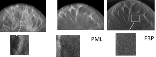
Besides novel acquisition strategies, another avenue that is being explored is improved tomographic reconstruction methods. Conventionally used linear image reconstruction methods like filtered back projection yields noisy image when a low dose acquisition protocol is used. We are investigating utility of statistical iterative reconstruction methods to mitigate this problem. These advanced image reconstruction methods allow modeling of imaging physics allowing for reduced noise images that have shown to improve detectability in tomographic imaging.
We use rigorous computer simulations and work closely with our clinical collaborators (Dr. Gary Whitman and Dr. Bill Geiser) are M. D. Anderson Cancer Center to investigate these aspects.
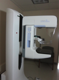
Hologic Dimensions DBT unit at M. D. Anderson Cancer center (above).
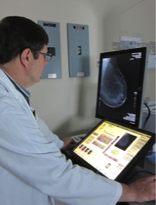
Dr. Geiser acquiring a DBT image (below)
Rigorous computer simulations showing comparison of DBT images with an iterative penalised maximum likelihood reconstruction (PML) and filtered back projection (FBP) reconstruction.
Combining Light/Radiation and Acoustics: Radiological (x-ray/gamma), optical and ultrasound imaging methods employed for in-vivo imaging of cancers or other malignancies have limitations when used as stand alone modalities. Our group is interested in modulating the effect of each one of these or combining the advantages offered by these modalities for efficient and novel detection schemes.
EM Wave Propagation in Biological Tissue: We use a combination of experiments, Monte Carlo methods and analytical methods to understand the forward propagation of EM waves (ionizing and non-ionizing radiation) in various tissue structures. This approach helps us with proving a feedback mechanism based design of efficient imaging techniques.
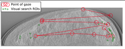
Understanding human perception - detection of low contrast signals in a complex background (collaborative project with Dr. Howard Gifford, Biomedical Engineering, UH)
Fig. (left) shows eye-tracking data (ASL eye tracking) of human observer and comparison with visual search observer tracking for a DBT image
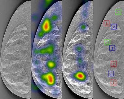
Fig. (left) shows Heat maps of two observer gaze on DBT image (SMI gaze tracking)
Psychophysics, Image Perception and Pre-Clinical Optimization: Our goal of devising methodologies for reduced dose imaging makes it imperative that we understand the signal detection and perception science involved in radiology. Psychophysical methods using human observer studies are the gold standard for optimizing various acquisition and reconstruction methods. These can be extremely time consuming and expensive. A mathematical model that mimics the human vision and perception will speed up such optimization techniques. In a collaborative project with the Biomedical Engineering department and radiologists at M. D. Anderson, we are developing a robust and clinically relevant mathematical model observer for tomographic breast imaging.
Innovative aspects of this research include the utility of a “visual search” mathematical model observer and use of eye tracking equipment to understand human perception of complex images and signal detection in complex backgrounds.
VIRTUALCLINICAL TRIALS FOR IMAGING PLATFORMS
- Computer simulation platforms to support clinical trials for novel imaging techniques.